Anchorage’s Experienced, High-Tech Obstetric Ultrasound.
Introduced in the 1950s, ultrasound imaging is very useful as a diagnostic tool in the field of obstetrics. Using sound waves instead of harmful ionizing radiation, ultrasound provides direct images of — and precise information about — both the developing fetus and the pregnancy.
Ultrasound: Monitoring And Measuring For Optimal Obstetric Care.
Ultrasound produces images by interpreting the echoes of high-frequency sound as it passes through different types of tissue. The various tissue types affect the rate of echo, which a computer translates into distinguishable images. These images are used to monitor and measure a variety of different aspects of pregnancy and fetal development, making ultrasound a vital — and indispensable — part of obstetric care today.
Comprehensive Obstetric Ultrasound Exams For Mother And Baby.

At Alaska Family Sonograms, our highly trained, credentialed and experienced sonographers are extremely skilled in obstetric ultrasound, which plays an essential role in the care of every pregnant woman. Combining our own capabilities with state-of-the-art ultrasound technology and an unsurpassed commitment to compassion, we perform ultrasound exams to:
At Alaska Family Sonograms, our highly trained, credentialed and experienced sonographers are extremely skilled in obstetric ultrasound, which plays an essential role in the care of every pregnant woman. Combining our own capabilities with state-of-the-art ultrasound technology and an unsurpassed commitment to compassion, we perform ultrasound exams to:
- Diagnose and confirm pregnancy early on – We can see the gestational sac, the yolk sac and the embryo by 5½ weeks. Our sonographers also confirm that the site of the pregnancy is within the uterine cavity.
- Document the viability of the fetus in the presence of vaginal bleeding in early pregnancy.
- Diagnose ectopic and molar pregnancies in the first trimester
- Help determine date of conception and delivery
- Assess and measure:
- The baby’s head and spine
- Fetal organs, including heart, stomach, bladder, kidneys, intestine.
- The baby’s extremities – arms, hands, fingers, legs, feet and toes
Your baby’s face - Detect, diagnose and measure fetal malformations.
- Determine gender, if desired, usually at 16 to 20 weeks.
- Assess the placenta – detect or exclude placenta previa, and evaluate for placental abnormalities in the presence of diabetes, RH isoimmunization and intrauterine growth restriction.
- Measure and monitor multiple pregnancies (twins and high-order multiples)
Detailed Fetal Anatomy Exam (CPT 76811)

A detailed fetal anatomy exam (sometimes called a level 2 exam) of the fetus may be requested when a pregnancy involves increased risk factors. These exams require a more in-depth look at the fetus and are recommended to be scheduled at 20 weeks. Our detailed fetal anatomy scans are read and interpreted by our radiologist Anthony Filly, MD. who specializes in high risk OB.
Fetal Echocardiography (CPT 76825, 76827, 93325)
Fetal echocardiograms focus solely on evaluating the structure and function of the fetal heart. These may be indicated if certain factors increase the risk of the fetus having heart problems. We perform these exams with pediatric cardiologists from Pediatric Cardiology of Alaska
Nuchal Translucency Screening (CPT 76813)
A nuchal translucency scan is performed between 11 and 13 weeks. This ultrasound is combined with a blood test (blood is retrieved with a finger poke, not a needle). The result gives the risk of a fetus having specific chromosomal abdnormalities ( Trisomy 13, 18, and 21).
Advanced, Experienced And Highly Compassionate Ultrasound Care.
In addition to regularly scheduled obstetric ultrasounds, Alaska Family Sonograms performs specialized ultrasound exams as needed. And just as important as our state-of-the-art capabilities and experienced skill, we provide the compassion, individualized attention, heartfelt understanding and support pregnant women and couples depend on. Our sonography team is well known in Anchorage for treating everyone like family, catering to their comfort and genuinely being there for them, whatever their needs.
State-Of-The-Art Diagnostic And 3D/4D Ultrasound In Anchorage.
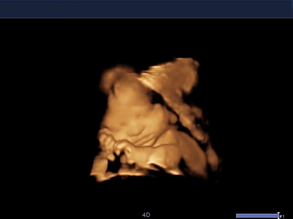
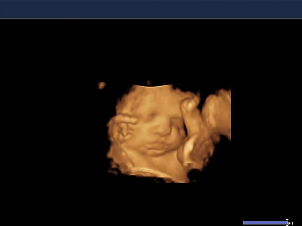
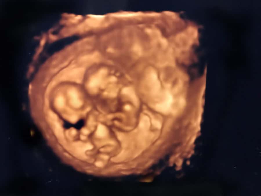
Also, a longstanding reference site for ultrasound leader Canon Medical (previously Toshiba), AFS has the most up-to-date technology, producing top-quality images for diagnostic purposes but, also, for 3D/4D imaging. We include 3D/4D imaging and pictures as a part of all of our exams starting in the late first trimester. 3D/4D ultrasound – particularly with our highly advanced equipment — provides truly spectacular real-time, three-dimensional footage of your baby, letting you “see” your baby up close even before your delivery date.

For more about obstetric ultrasound at Alaska Family Sonograms in Anchorage, or to schedule an appointment, call 907.561.3601. You can also request an appointment using our easy online form.
FAQ
Absolutely. We have introduced many parents to their unborn child through the use of sonographic images and video. You will receive printed images and you will be sent a link (via text or email whichever you prefer) that you can access your exam at as well. We recommend you access your exam from the link and download the images/video so you can permanently keep them as the link will remain active for two months.
The gender of your baby will typically be determined during a routine anatomy scan. This is performed around 18-20 weeks gestational age.
We do not perform an exam only to determine a baby’s gender. We feel every baby deserves a complete and thorough sonogram.
If you do not want to know the gender please let the sonographer know before your exam begins.
If you are wanting a copy of your exam images/video we can send you another link to access them, just give us a call to request this.
The duration of the exam will vary based on what specific type of OB exam was requested. The typical duration will be around 45 minutes.
Nothing is specifically required for these exams however we do recommend you have something to eat prior to your appointment. This can sometimes help wake baby up. Having a baby awake and moving can help us obtain the images we need for your exam.
